Newly added to the STAT!Ref Collection are two new Opthalmology resources from Oxford University Press, Inc. As a reminder, All STAT!Ref content is full-text, cross-searchable and every single resource is available on the STAT!Ref Mobile App.
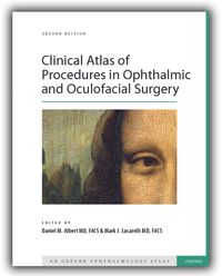
The second edition of Clinical Atlas of Procedures in Ophthalmic and Oculofacial Surgery provides an overview of a broad range of contemporary, well-established, and accepted ophthalmic surgical procedures with clear illustrations of surgical fundamentals that cover key intraoperative and postoperative points.
This new edition of the Atlas includes streamlined, more uniform chapters, book-ended by detailed and instructive tables of indications and complications. More than 2,500 detailed, professionally-rendered line drawings and full-color photographs supplement succinct information on surgical procedures. The high-quality illustrations and images are laid out in a fluid design to help the reader quickly pinpoint the fundamentals of each procedure.
With innovations and techniques frequently evolving ophthalmic surgery, the second edition of Clinical Atlas of Procedures in Ophthalmic and Oculofacial Surgery provides the clear and comprehensive platform needed to navigate the fast-moving field of surgical ophthalmology, and will surely continue to prove useful to the trainee, the ophthalmologist, the teacher, and, most importantly, to the patients whom they ultimately serve.
Overview:
- Doody's Core Title
- Richly illustrated, with more than 2,500 clear line drawings and high-quality, full-color surgical photographs
- Comprehensive text covering topics ranging from introduction to surgical tools and basic surgical technique to complex surgical reconstructions
- Contributions from over 150 internationally recognized experts for trusted insight into advanced surgical technique and new developments within the field
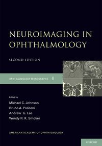
Since the publication of the original edition of this American Academy of Ophthalmology Monograph in 1992, new techniques and special sequences have improved our ability to detect pathology in the orbit and brain that are significant for the ophthalmologist. In this second edition of Neuroimaging in Ophthalmology, Johnson, Policeni, Lee, and Smoker have updated the original content and summarized the recent neuroradiologic literature on the various modalities applicable to CT and MR imaging for ophthalmology.
The goal of this resource is to reinforce the critical importance of accurate, complete, and timely communication–from the prescribing ophthalmologist to the interpreting radiologist–of the clinical findings, differential diagnosis, and presumed topographical location of the suspected lesion in order for the radiologist to perform the optimal imaging study, and ultimately, to receive the best interpretation.
Overview:
- Doody's Core Title
- Only resource to summarize the recent neuroradiologic literature on the various modalities applicable to CT and MR imaging for ophthalmology
- Emphasizes vascular imaging advances (e.g., MR angiography (MRA), CT angiography (CTA), MR venography (MRV), and CT venography (CTV) and specific MR sequences (e.g., fat suppression, fluid attenuation inversion recovery (FLAIR), gradient recall echo imaging (GRE), diffusion weighted imaging (DWI), perfusion weighted imaging (PWI), and dynamic perfusion CT (PCT))
- Includes tables that outline the indications, best imaging recommendations for specific ophthalmic entities, and examples of specific radiographic pathology that illustrate the relevant entities
Bundle these resources with The Wills Eye Manual: Office and Emergency Room Diagnosis and Treatment of Eye Disease.
Have any questions about these new resources? Please call 800-901-5494 or fill out this form to speak with a STAT!Ref Team Member today.

